TOPIC 13: Systems Physical Assessment IV
Introduction:
The male reproductive system is anatomically divided into external and internal genital organs. The penis and scrotum are external organs and are easily inspected and palpated, whereas the internal structures have limited accessibility. So, the assessment includes both the male reproductive system and the urinary system.
The musculoskeletal system provides shape and support to the body, allows movement, protects the internal organs, produces red blood cells in the bone marrow and stores calcium and phosphorus in the bones. This chapter also presents the sensory-neurologic assessment, including information on assessing cerebral function, cranial nerves (CNs), sensation, and reflexes. The neuromuscular function, just one aspect of the highly complex neurologic system. The goal of overall physical assessment is to detect risk factors, potential problems, or any dysfunction early and then to plan appropriate interventions, including teaching health promotion and disease prevention and implementing treatment measures.
Topic Learning Outcome (TLOs):
By the end of this topic, you should be able to:
- Demonstrate a physical assessment of the male genitourinary system
- Document male genitourinary findings
- Demonstrate a physical assessment of the motor-musculoskeletal system
- Document motor-musculoskeletal findings
- Demonstrate a physical assessment of the sensory-neurologic system
- Document sensory-neurologic system findings
MALE GENITO-URINARY
In adult men, a complete examination includes assessment of the external genitals and prostate gland, and for the presence of any hernias. Nurses in some practice settings performing routine assessment of clients may assess only the external genitals. The male reproductive and urinary systems share the urethra, which is the passageway for both urine and semen. Therefore, in physical assessment of the male these two systems are frequently assessed together.

Performing the Male Genitals and Inguinal Physical Assessment
The techniques of inspection and palpation are used to examine the male genitals. The table below describes how the nurse can assess the male genitals and inguinal area.


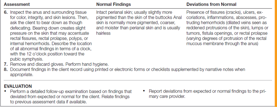
Performing the Anus Physical Assessment
For the anal examination, an essential part of every comprehensive physical examination, involves only inspection. The table below describes how to assess the rectum and anus.


Documentation
Penis normal shape without lesions. Testicles normal shape and contour without tenderness. Epididymides normal shape and contour without tenderness. No discharge or hernias. Rectum normal tone to sphincter. Prostate normal shape and contour without nodules. Stool hemoccult negative. No external hemorrhoids. No skin lesions.
For better understanding and practice, you may watch the following videoclip on “Physical Examination of the Male Genitalia, Hernias, Anus, and Rectum”
Video:MUSCULOSKELETAL SYSTEM
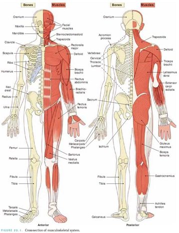
Before beginning your assessment, you need to understand how the musculoskeletal system works. It consists of three major components: bones, muscles, and joints. Tendons, ligaments, cartilage, and bursae serve as connecting structures and complete the system. Diagram below illustrates the musculoskeletal system, anterior and posterior view.
The nurse usually assesses the musculoskeletal system for muscle strength, tone, size, and symmetry of muscle development, and for tremors. A tremor is an involuntary trembling of a limb or body part. Tremors may involve large groups of muscle fibers or small bundles of muscle fibers. An intention tremor becomes more apparent when an individual attempts a voluntary movement, such as holding a cup of coffee. A resting tremor is more apparent when the client is relaxed and diminishes with activity. A fasciculation is an abnormal contraction of a bundle of muscle fibers that appears as a twitch.
Bones are assessed for normal form. Joints are assessed for tenderness, swelling, thickening, crepitation (a crackling, grating sound), and range of motion. Body posture is assessed for normal standing and sitting positions.
Performing the Musculoskeletal Physical Assessment
The table below describes how to assess the musculoskeletal system.


Documentation
· Both extremities are equal in size.
· Have the same contour with prominences of joints.
· No involuntary movements.
· No edema
· Color is even.
· Temperature is warm and even.
· Has equal contraction and even.
· Can perform complete range of motion.
· No crepitus must be noted on joints.
· Can counter act gravity and resistance on ROM.For better understanding and practice, you may watch the following videoclip on “Physical Examination of the Musculoskeletal System”
Video:Sensory-Neurologic System
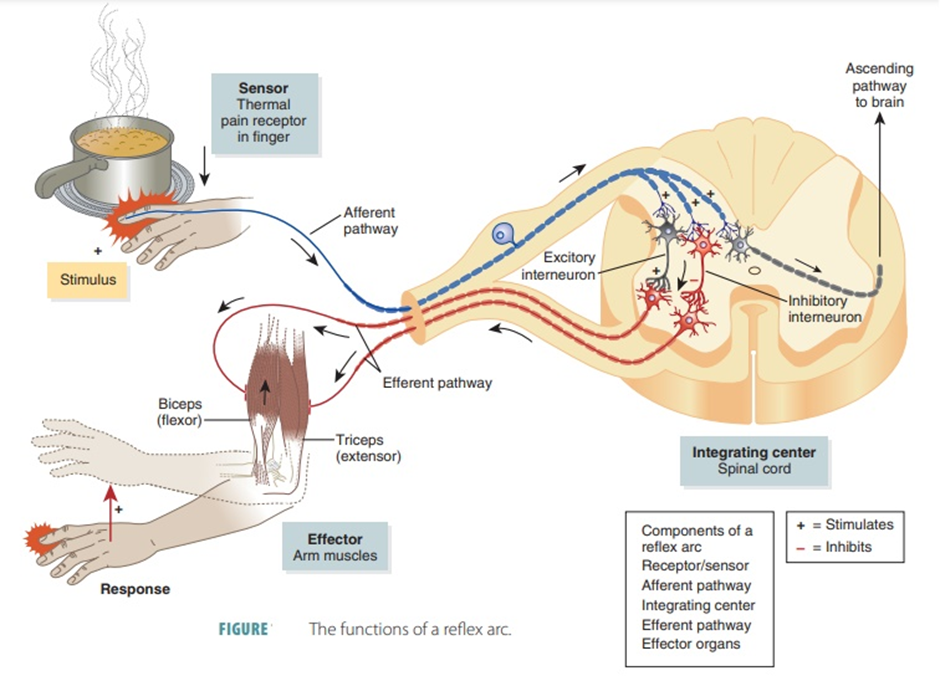
Sensory impulses are transmitted to the brain through afferent, or ascending, pathways. Motor impulses are transmitted from the brain to muscles through the efferent, or descending, pathways as shown in the following diagram.
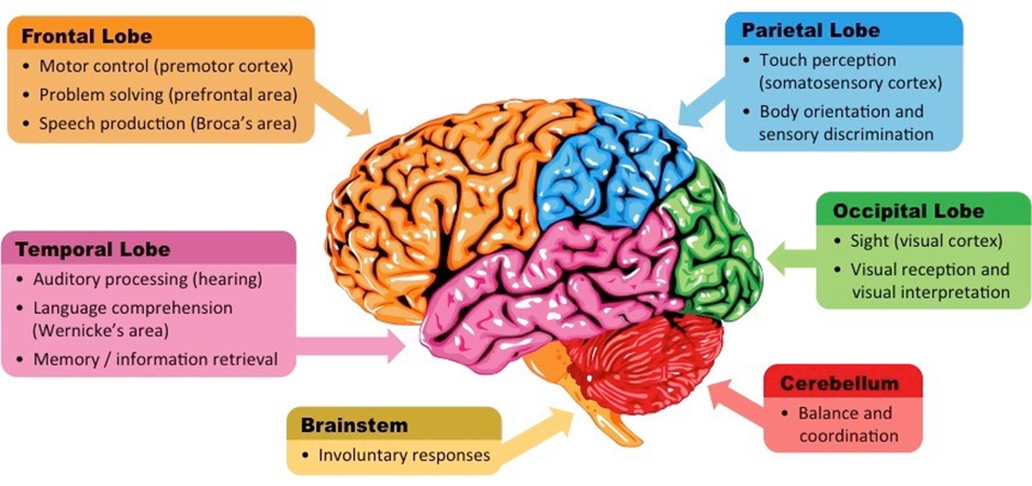
The brain is composed of gray matter, made up of neuronal cell bodies, and white matter, composed of axons and dendrites. The brain consists of four major structures: the cerebrum, diencephalon, cerebellum, and brainstem. These and other components of the brain are shown in the below diagram.
There are 12 pairs of cranial nerves originate from the brain and are called the peripheral nerves of the brain. As shown in the following diagram.
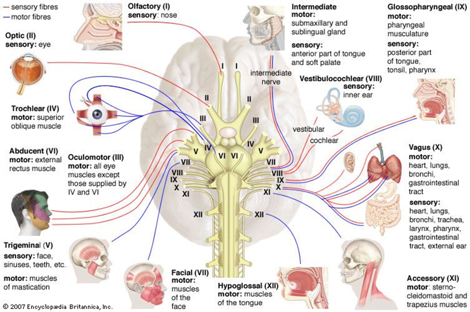
Branching from the spinal cord are 31 pairs of spinal nerves: 8 cervical, 12 thoracic, 5 lumbar, 5 sacral, and 1 coccygeal as shown in the following diagram.

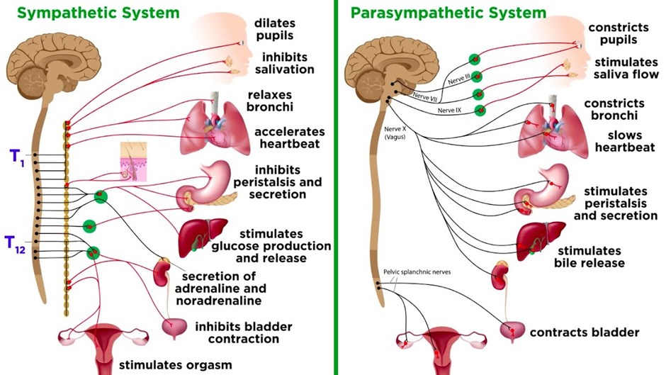
The autonomic nervous system is divided into the sympathetic and parasympathetic, with efferent fibers to muscle, organs, or glands. Usually, the two systems work opposite each other. The sympathetic allows the body to respond to stressful situations. The parasympathetic functions when all is normal. The sympathetic nerves exit the spinal cord between the level of the first thoracic and the second lumbar vertebrae. The preganglionic nerves descend the cord and exit, and then enter a relay station known as the sympathetic chain. The impulse is then transmitted to a postganglionic neuron that goes to the target organ to stimulate a response. The following diagram illustrates the autonomic nervous system.
Performing the Neurologic Assessment
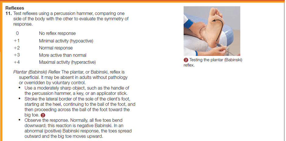
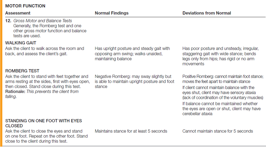
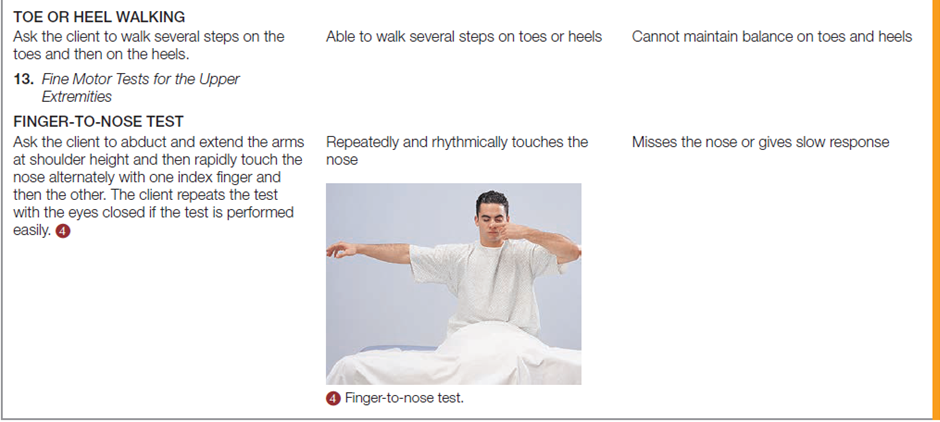
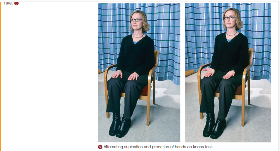
The table below describes how to assess the neurologic system.






Documentation
Alert and oriented to situation, person, place, and time. Behavior appropriate to situation and developmental age. Clear speech and follow verbal commands. Cranial nerves II to XII grossly intact. Pupils Equal, Round, React to Light and Accommodation (PERRLA). Active range of motion all extremities with symmetry strength. Peripheral sensation intact.
For better understanding and practice, you may watch the following videoclip on “Physical Examination of the Nervous System: Sensory System and Reflexes”
Video: