TOPIC 12: Systems Physical Assessment III
Introduction:
The assessment of the genitourinary (GU) system and breast are essential to obtaining a picture of a woman’s overall health status. Because most breast masses are detected by self-examination, teaching your patients how to examine their breasts is just as critical and the female reproductive systems undergo cyclical changes in response to hormonal levels throughout the life cycle.
The stomach, small and large intestines, liver, gallbladder, pancreas, spleen, kidneys, ureters, bladder, aortic vasculature, spine, uterus and ovaries, or spermatic cord are all located in the abdomen. So, abdominal assessment provides information about a variety of systems can also provide vital information about the health status of every other system.
Topic Learning Outcome (TLOs):
By the end of this topic, you should be able to:
- Demonstrate a physical assessment of the breast.
- Document breast findings.
- Demonstrate an abdominal physical assessment.
- Document abdominal assessment findings.
- Demonstrate a physical assessment of the female genitourinary system.
- Document female genitourinary findings
BREASTS AND AXILLAE
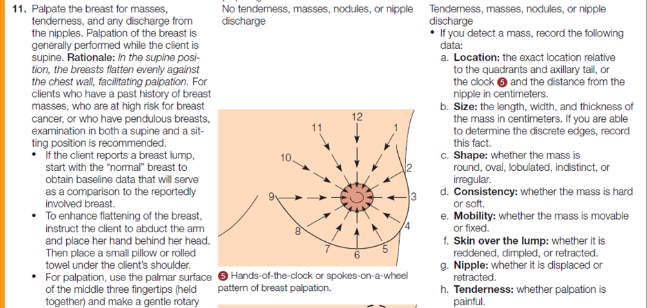
The breasts of men and women need to be inspected and palpated. Men have some glandular tissue beneath each nipple, a potential site for malignancy, whereas mature women have glandular tissue throughout the breast. In females, the largest portion of glandular breast tissue is located in the upper outer quadrant of each breast. A projection of breast tissue from this quadrant extends into the axilla, called the axillary tail of Spence, as shown in the following diagram.

The majority of breast tumors are located in this upper outer breast quadrant including the tail of Spence. During assessment, the nurse can localize specific findings by dividing the breast into quadrants and the axillary tail.
Performing the Breast and Axillae Physical Assessment
The table below describes how to assess the breast and axillae.




Documentation
Inspection of the Breast
- The overlying the breast should be even.
- May or may not be completely symmetrical at rest.
- The areola is rounded or oval, with same color.
- Nipples are rounded, everted, same size and equal in color.
- No “orange peel” skin is noted which is present in edema.
- The veins maybe visible but not engorge and prominent.
- No obvious mass noted.
- Not fixated and moves bilaterally when hands are abducted over the head, or is learning forward.
- No retractions or dimpling.
Palpation of the Breast
- No lumps or masses are palpable.
- No tenderness upon palpation.
- No discharges from the nipples.
For better understanding and practice, you may watch the following videoclip on “Physical Examination of the Breast and Axillae”
Video:
ABDOMEN
The nurse locates and describes abdominal findings using two common methods of subdividing the abdomen: quadrants and regions. To divide the abdomen into quadrants, the nurse imagines two lines: a vertical line from the xiphoid process to the pubic symphysis, and a horizontal line across the umbilicus, as following diagram.

Using the second method, division into nine regions, the nurse imagines two vertical lines that extend superiorly from the midpoints of the inguinal ligaments, and two horizontal lines, one at the level of the edge of the lower ribs and the other at the level of the iliac crests, as shown in the following diagram.
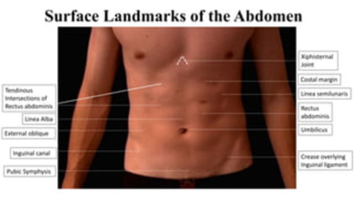
In addition, practitioners often use certain landmarks to locate abdominal signs and symptoms. These are the xiphoid process of the sternum, the costal margins, the anterosuperior iliac spine, the umbilicus, the inguinal ligaments, and the superior margin of the pubic symphysis, as in the following diagram.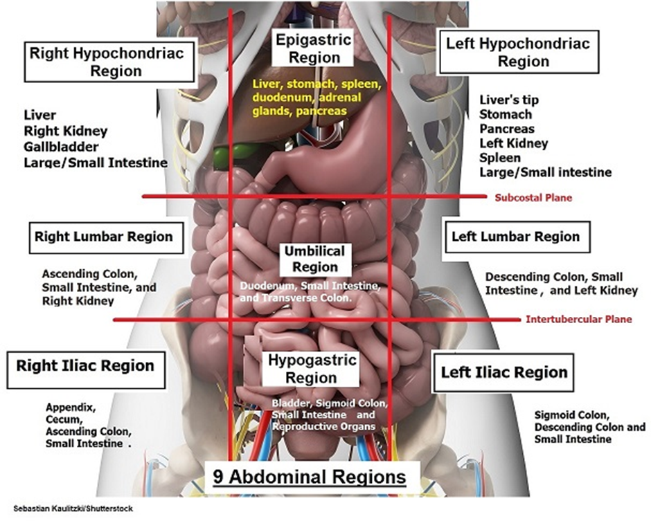
Performing the Abdomen Physical Assessment
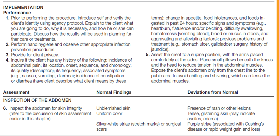
Assessment of the abdomen involves all four methods of examination (inspection, auscultation, palpation, and percussion). When assessing the abdomen, the nurse performs inspection first, followed by auscultation, percussion, and/or palpation. Auscultation is done before palpation and percussion because palpation and percussion cause movement or stimulation of the bowel, which can increase bowel motility and thus heighten bowel sounds, creating false results.
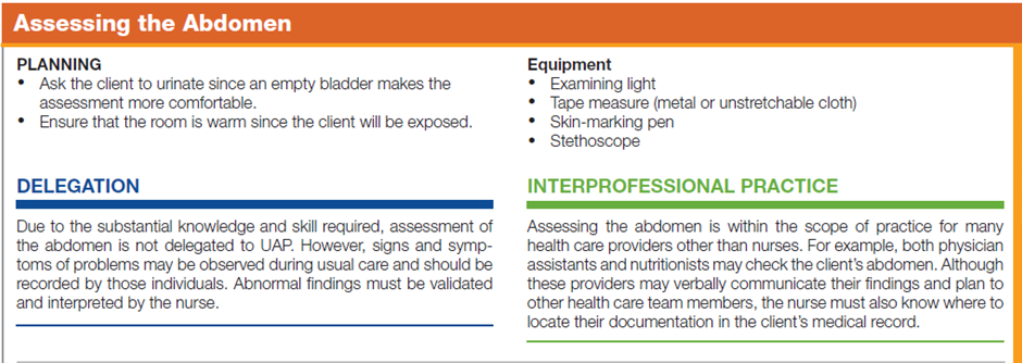
The table below describes how to assess the abdomen.


Documentation
The abdomen is the same color as the rest of the body with sparse hair distribution close to the perineum. The abdomen is rounded, symmetrical with straie around the waist. The umbilicus is midline and no odor or discharge noted. The abdomen is soft, non-distended, non-tender with positive bowel sounds to all four quadrants and no guarding noted during the assessment. No distention to the suprapubic area and no edema to face, hand, or lower extremities noted.
For better understanding and practice, you may watch the following videoclip on “Physical Examination of the Abdomen”
Video:FEMALE GENO-URINARY
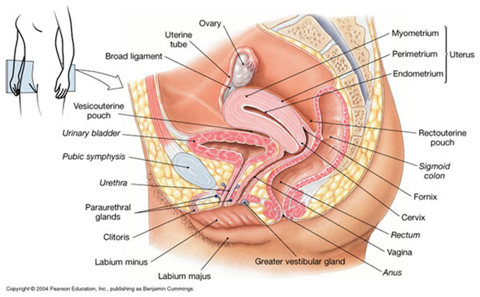
The system consists of external and internal genitalia, which develop and function according to the hormonal influences that affect fertility and childbearing. It also consists of urinary structures. External genitalia include the mons pubis, clitoris, vestibule, labia majora, labia minora, vaginal introitus, hymen, Bartholin’s glands, Skene’s glands, and the urethral meatus. Internal genitalia include the vagina, cervix, uterus, adjacent structures (adnexa), ovaries, and fallopian tubes. Internal urinary structures include the ureters, bladder, and urethra, as in the following diagram.

Performing the Female Genitals and Inguinal Physical Assessment
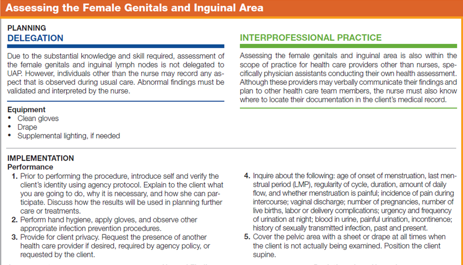
The examination of the genitals and reproductive tract of women includes assessment of the inguinal lymph nodes and inspection and palpation of the external genitals. Completeness of the assessment of the genitals and reproductive tract depend on the needs and problems of the individual client. In most practice settings, generalist nurses perform only inspection of the external genitals and palpation of the inguinal lymph nodes. Examination of the genitals usually creates uncertainty and apprehension in women, and the lithotomy position required for an internal examination can cause embarrassment.
The table below describes how to assess the female genitals and inguinal area.


Documentation
External genitalia without erythema, exudate or discharge. Vaginal vault is without discharge. Cervix is of normal color without lesion. The os is closed. There is no bleeding noted. Uterus is noted to be of normal size and nontender. No cervical motion tenderness is seen. No masses are palpated. The adnexa are without masses or tenderness.
For better understanding and practice, you may watch the following videoclip on “Physical Examination of the Female Genitalia, Anus and Rectum”
Video: